![]()
Sierra College
Biological Sciences'
Microscopic Slide Collection and Associated Images
Trypanosoma lewisi (w.m.)
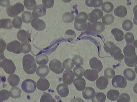
Photomicrograph of Trypanosoma lewisi (four cells) in a blood smear. Magnification: 1000x.
A photomicrograph of a prepared slide showing Trypanosoma lewisi in a blood smear, magnified 1000x. The four trypanosomes appear as thin, eel-like cells surrounded by disc-shaped erythrocytes (RBCs). Each trypanosome has a dark, oval-shaped nucleus and a smaller, dark, spherical kinetoplast. A single flagellum extends the length of the cell (or beyond), attached to it by an undulating membrane that appears fin-like.
Copyright 2014 Sierra College Biological Sciences Department
Last updated: October 15, 2014
![]()
![]()