![]()
Sierra College
Biological Sciences'
Microscopic Slide Collection and Associated Images
Euglena (w.m.)
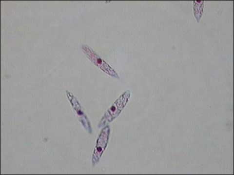
Photomicrograph of a stained preparation of Euglena cells. Nuclei stain as dark spots, flagella are not readily visible. Magnification: 400x.
A photomicrograph of a prepared slide showing Euglena stained pink and magnified 400x. These single-celled algae are usually linear, but lack cells walls so are able to change their shape. The dark, centrally located, oval-shaped structure visible within each cell is the nucleus. Each cell also has a single flagellum extending from one end, but these are very thin and often difficult to observe.
Copyright 2014 Sierra College Biological Sciences Department
Last updated: October 15, 2014
![]()
![]()