![]()
Sierra College
Biological Sciences'
Microscopic Slide Collection and Associated Images
Euglena (w.m.)
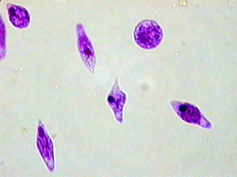
Photomicrograph of Euglena, showing variation in cell shape. Magnification: 400x.
A photomicrograph of a prepared slide showing Euglena stained pink and magnified 400 times. The dark pink ovals visible within the cells are nuclei. Each cell has one flagellum extending from the more rounded end, but these are not visible on all of the cells present. These organisms do not have rigid cell walls, so can take on a variety of shapes.
Copyright 2014 Sierra College Biological Sciences Department
Last updated: October 15, 2014
![]()
![]()